Review Article
말초동맥질환 환자의 심장재활
이종화1,*
Cardiac Rehabilitation for the Patients with Peripheral Artery Disease
Jong Hwa Lee1,*
1Department of Physical Medicine and Rehabilitation, Dong-A University College of Medicine, Busan 49201, Korea
*교신저자 : 이종화, 49201 부산광역시 서구 대신공원로 26 동아대학교 의과대학 재활의학교실 / 동아대학교병원 심뇌혈관센터, Tel : +82-51-240-5690, Fax : +82-51-254-8511, e-mail :
jhlee08@dau.ac.kr
© Copyright 2021 The Korean Space Science Society. This is an Open-Access article distributed under the terms of the
Creative Commons Attribution Non-Commercial License (http://creativecommons.org/licenses/by-nc/4.0/) which permits
unrestricted non-commercial use, distribution, and reproduction in any
medium, provided the original work is properly cited.
Received: May 21, 2021; Accepted: Jun 11, 2021
Published Online: Jun 30, 2021
Abstract
Patients with peripheral artery disease (PAD) usually have functional impairment, faster functional decline, and poorer quality of life (QoL). It is important to treat PAD properly because it can cause of increasing risk of cardiovascular diseases and related mortality. In addition to anti-platelet agents and statins, cardiac rehabilitation including lifestyle modification and aerobic exercise therapy must be provided to these patients. The mechanism leading to exercise induced improvement may include changes in the way that oxygen is used by exercising the muscle, improved endothelial function, reduced cardiovascular disease risk factors and improved gait as well as increased blood supply. Prior to prescribing exercise, treadmill exercise teat may be given priority to assess functional status. Structured supervised treadmill exercise and home-based exercise each significantly improve functional status and QoL and reduce leg symptoms. In patients with claudication, alternative strategies of exercise, including upper-body ergometry, cycling, and pain-free walking can be beneficial to improve walking ability and functional status. Studies with long-term follow-up have demonstrated a persistent benefit of those exercises in patients with claudication. Furthermore, the risk-benefit ratio is favorable, with an excellent safety profile in patients screened for absolute contraindications to exercise. In order to increase the effectiveness of exercise therapy and prevent problems, it will be most important to fully evaluate the patient’s condition and prescribe appropriate exercise intensity, duration, and modalities.
Keywords: Peripheral artery disease; Intermittent claudication; Exercise therapy; Quality of life
서론
말초동맥질환(peripheral artery disease, PAD)은 뇌와 심장으로 가는 주요 혈관 이외의 다른 혈관들이 비정상적으로 좁아지면서 발생하는 질환을 일컫는다. 대개 동맥 경화나 염증 반응에 의하여 색전, 혈전이 생성되거나 협착되어 혈관의 기능과 구조에 이상을 초래하게 된다. 50세 미만에서는 드물지만 이후 연령대에서부터는 뚜렷한 증가세가 관찰되어, 65세 이상에서는 약 10-15%, 80세 이상에서 약 20% 이상의 발생률이 보고되고 있다[1]. 고령을 포함하여 흡연, 당뇨, 고혈압, 이상지질혈증 등이 유발의 주된 원인으로 간주된다[2]. 임상 양상은 무증상에서부터 궤양, 괴저 발현 등 다양하나 가장 특징적으로 간헐적 파행(intermittent claudication)이 나타날 수 있다. 보행을 하거나 운동을 할 때 다리에 쥐가 나거나 통증, 피로감 등을 호소하지만 활동을 멈추고 쉬면 곧바로 증상이 소실되는 특징을 보인다. 말초동맥질환은 심혈관계 질환에 대해 독립적인 위험인자로 알려져 있으며, 이와 같은 혈관 문제를 가진 환자는 그렇지 않은 사람들에 비해 심혈관계 질환 발생률과 사망률이 2-3배 이상 높을 수 있다[3]. 사지를 절단할 정도로 악화된 경우에는 5년 생존률이 30% 미만을 보일 정도로 예후가 좋지 않다. 또한 이동 능력의 소실과 같은 기능적 손상이 점차 증가하게 되면서 삶의 질이 떨어지게 되는 결과를 경험하게 된다[4]. 이러한 위험을 줄이기 위한 치료로 우선 적절한 항혈전제와 스타틴 제재를 사용해야 한다. 반드시 금연을 해야하며, 항고혈압제를 통해 안정적으로 혈압을 관리해야 한다[5]. 그리고 무엇보다 지속적인 유산소 운동을 포함하는 심장재활을 시행하는 것이 중요하다. 감독 하 운동 치료(supervised exercise therapy) 혹은 가정 기반의 운동 치료(home-based exercise therapy)는 모두 보행 능력, 전반적인 기능 상태, 그리고 건강관련 삶의 질을 향상시키는데 매우 중요한 역할을 하는 것으로 알려져 있다[6]. 질환의 특성 상 운동 능력이 저하된 환자들에서 적절한 강도와 간격으로 운동을 지속적으로 시행하는 것은 매우 중요하면서도 임상적으로 적용하기 어려운 한계도 존재한다. 지금부터 기존의 연구들을 바탕으로 말초동맥질환 환자에서 적용할 수 있는 심장재활 운동 방법에 대해 살펴보도록 하겠다.
본론
말초동맥질환 환자들에서 운동 치료를 시행하는 궁극적인 목표는 일상 생활을 정상적으로 영위하기 위해 필요한 운동 수행 능력을 향상시켜 삶의 질을 높이고 전반적인 기능 상태를 호전시키는데 있다. 운동 치료의 효과를 판단하는데 있어 하지의 혈역동학적 평가보다 트레드밀 운동 검사, 6분 보행 검사, 그리고 환자 관련 삶의 질 평가 도구 등을 우선적으로 사용하는 것은 이러한 기능적인 영역을 반영할 수 있기 때문이다.
1) 운동을 통한 기능 향상의 기전(mechanism)
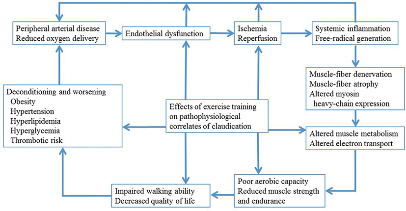
운동으로 인한 기능 향상의 기전에 대해서 완전하게 알려져 있지는 않으나, 산소가 근육을 운동시키는데 사용되고 혈관 내피 기능을 호전시키며, 심혈관계 질환의 위험인자들을 줄이면서 혈액 공급을 증가시키는 등의 역할을 수행하기 때문일 것으로 예상하고 있다. 최근에 제안되는 기전들로 말초동맥질환을 가진 환자들에 대해 규칙적인 운동을 하는 것이 장애의 고리(cycle of disability)를 깨는 것과 연관이 있을 것으로 여겨지고 있다(Fig. 1) [7].
2) 구조화된 운동 치료(structured exercise therapy)의 권고(recommendation) [
8]
잘 구조화된 운동 치료는 말초동맥질환 환자의 치료에 매우 중요한 요소이다. 해당 프로그램의 구성 요소들에 대해 Table 1과 2에 제시하였다. 파행에 대한 최초 치료방법으로서 감독 하 운동 프로그램의 효과를 증명하는 연구들은 계속 발전하여 설득력을 유지해오고 있다[9]. 18개월부터 7년에 이르는 장기간 경과 관찰한 연구들에서도 파행을 보이는 환자들에서 감독 하 운동의 지속되는 이점들을 뒷받침하고 있다[10,11]. 말초동맥질환에 대한 감독 하 운동의 위험-이익 상관관계에서 긍정적인 결과가 관찰되었고, 운동에 대한 절대 금기인 운동-제한 심혈관 질환, 절단 혹은 휠체어 생활, 그리고 운동을 방해할 수 있는 다른 주요 동반질환들을 잘 차단했을 경우 안전성도 매우 높은 것으로 보고되었다[12]. 운동 기능은 4-8주부터 점진적으로 좋아지기 시작해서 12-24주를 넘어서까지 지속적으로 향상된다. 병원에서의 감독 하 운동이 가정에서의 운동에 비해 보행 거리의 호전에 있어 더 효과적이나 삶의 질이나 자가 보고식 기능의 호전에는 큰 차이를 보이지 않았다[13].
Table 1.
Structured Exercise Program for Peripheral Artery Disease
|
Structured exercise program |
Supervised exercise therapy (COR I, LOE A)
• Program takes place in a hospital or outpatient facility.
• Program uses intermittent walking exercise as the treatment modality.
• Program can be standalone or within a cardiac rehabilitation program.
• Program is directly supervised by qualified healthcare provider(s).
• Training is performed for a minimum of 30–45 min/session; sessions are performed at least 3 times/week for a minimum of 12 weeks.
• Training involves intermittent bouts of walking to moderate-to-maximum claudication, alternating with periods of rest.
• Warm-up and cool-down periods precede and follow each session of walking. |
Structured community- or home-based exercise program (COR IIa, LOE A)
• Program takes place in the personal setting of the patient rather than in a clinical setting.
• Program is self-directed with guidance of healthcare providers.
• Healthcare providers prescribe an exercise regimen similar to that of a supervised program.
• Patient counseling ensures understanding of how to begin and maintain the program and how to progress the difficulty of the walking (by increasing distance or speed).
• Program may incorporate behavioral change techniques, such as health coaching or use of activity monitors |
Download Excel Table
Table 2.
Recommendations for Structured Exercise Therapy for Peripheral Artery Disease
|
COR |
LOE |
Recommendations |
|
I |
A |
In patients with claudication, a supervised exercise program is recommended to improve functional status and QOL and to reduce leg symptoms. |
|
I |
B-R |
A supervised exercise program should be discussed as a treatment option for claudication before possible revascularization. |
|
IIa |
A |
In patients with PAD, a structured community- or home-based exercise program with behavioral change techniques can be beneficial to improve walking ability and functional status. |
|
IIa |
A |
In patients with claudication, alternative strategies of exercise therapy, including upper-body ergometry, cycling, and pain-free or low-intensity walking that avoids moderate-to-maximum claudication while walking, can be beneficial to improve walking ability and functional status. |
Download Excel Table
3) 운동 프로그램을 위한 사전 평가
운동 기능 상태를 평가하기 위해 우선적으로 고려되는 것은 트레드밀 운동부하검사이다. 운동에 따른 환자의 심박동과 혈압 반응뿐 아니라 운동 중 파행의 역치를 포함한 정보를 얻을 수 있다. 이는 비정상적인 심장 반응이 발생할 수 있는 시점의 심박동 혹은 혈압에 도달하기 전에 다리 통증으로 인해 평가가 제한되는 것을 통해 감지할 수 있다. 훈련 이후 보행 능력의 향상에 의해 평가 중 드러나지 않았던 심장 증상들이 나타나는 경우도 종종 있음을 이해하고 있어야 한다. 부득이 트레드밀 검사가 어려운 경우 6분 보행 검사를 시행할 수 있지만, 운동 처방을 위해서는 상대적으로 효용성이 떨어지는 부분이 있다. 운동 검사를 시행할 때에는 발과 피부의 상태를 면밀히 관찰하여 평가 전후 상처가 발생하지 않도록 주의를 기울이는 것이 반드시 필요하다.
운동부하검사를 위해 여러 가지 프로토콜이 제시되고 있다. Gardner [14]가 제시한 단계적(graded) 프로토콜은 3.2 km/h 속도를 유지한 채로 경사도를 0%에서 매 2분마다 2%씩 증가시키는 방법으로 최대 30분간 시행할 수 있다. 변형 프로토콜으로는 2.0 km/h의 속도로 매 4분마다 경사도를 2% 증가시키거나, 4.4 km/h의 속도로 매 분마다 경사도를 2% 증가시키는 방법도 있다. Hiatt [15]가 제시한 프로토콜은 3.2 km/h 속도를 유지한 채로 경사도를 0%에서 매 3분마다 3.5%씩 증가시키는 방법이다. 그 밖에 경사도를 10-12%로 고정하거나 속도를 3 km/h로 고정한 채로 검사를 진행하는 고정식(fixed) 프로토콜도 제시된 바 있다. 검사 중 환자에게 일정한 간격(예, 30초 간격)으로 파행 통증 유무에 대해 질문하여야 한다. 대개 다리통증 척도(Table 3)를 사용하여 최초 파행 발생 시간과 더 이상 보행을 할 수 없는 최대 보행 가능 시간을 물어본다. 연구에 따르면 말초동맥질환의 상태 평가를 위해서는 단계적 프로토콜을 적용하여 최대 보행 시간을 측정하는 것이 가장 신뢰도가 높다고 알려져 있다[16].
Table 3.
Intermittent Claudication Rating Scale
|
Grade |
Symptoms |
|
0 |
No pain |
|
1 |
Initial, minimal pain |
|
2 |
Moderate pain, bothersome pain |
|
3 |
Intense pain |
|
4 |
Maximal pain, cannot continue |
Download Excel Table
4) 운동 프로그램 처방
운동을 통한 효과를 얻기 위해서는 한 세션 당 최소 30분 이상, 일주일에 최소 3 세션 이상의 훈련을 최소 12주 이상 지속하는 것이 필요하며 유산소 운동의 강도는 거의 중등도에서 최대 통증 수준에 가까이 가도록 하는 것이 권장된다. 운동 전 준비운동(warm-up)과 운동 후 정리운동(cool-down)은 각각 5분 이상씩 시행하여야 한다. 트레드밀을 이용한 일반적인 단계적 운동 방법은 Table 4에서 7에 각각 정리하였다[17,18].
Table 4.
Treadmill Exercise Program I
|
Exercise program |
• Start at 3.2 km/h speed and 0% grade.
• Use handrails only to keep balance, do not hold them too tight.
• Stop the treadmill completely when the pain scale reaches 2-3/4 points.
• Restart the exercise at the same intensity, after the discomfort disappears.
• Repeat rest/exercise sessions.
• Increase the load when walking is possible without leg discomfort for more than 8 min.
a) Increase the speed by 0.3 km/h when it is possible to walk for 8 min.
b) Increase the grade by 1% when it is possible to walk at 5.4 km/h or reach a speed that no longer can be raised. |
Download Excel Table
Table 5.
Treadmill Exercise Program II (including when CPET is not performed)
|
Exercise program |
• Walk at a comfortable speed and 0% grade to reach a lower extremity pain scale of 2-3/4 points after 5-10 min.
• When lower extremity pain develops, rest (in a sitting or standing position) until the pain disappears, and then repeat rest/exercise until the whole exercise time reaches about 60 min.
• Continue the exercise in a similar manner until the cumulative exercise time is approximately 30-45 min out of the total exercise time of 60 min.
• Increase the intensity gradually (0.2 km/h speed or 1% grade), when it is possible to walk continuously for more than 10 min.
• Continue the exercise when lower extremity pain reaches 2-3/4 points after 5-10 min and the cumulative exercise time reaches to 30-45 min during 60 min. |
Download Excel Table
Table 6.
Exercise Program if Lower Extremity Symptoms Occur in Non-treadmill Exercise Such as Lower Extremity Ergometer
|
Exercise program |
• Start at 50-80 revolutions per minute (RPM) until 2-3/4 points lower extremity pain develops after 5-10 min of exercise.
• When lower extremity pain develops, rest until the pain disappears, and then repeat rest/exercise until the whole exercise time reaches about 60 min.
• Continue the exercise in a similar manner until the cumulative exercise time is 30-45 min out of the total exercise time of 60 min.
• Increase the RPM little by little, when the exercise is consecutively possible over 10 min.
• Continue the exercise when lower extremity pain reaches 2-3/4 points after 5-10 min and the cumulative exercise time reaches to 30-45 min during 60 min. |
Download Excel Table
Table 7.
Exercise Program if Lower Extremity Symptoms do not Occur in Non-treadmill Exercise Such as Upper Extremity Ergometer
|
Exercise program |
• Start at a comfortable intensity around 50 revolutions per minute (RPM).
• Repeat 2 min exercise/2 min rest for 60 min.
• Gradually, increase exercise time, such as 5 min exercise/1 min exercise.
• Exercise 2 min exercise/2 min rest for 60 min
• Gradually, increase exercise time from 20 min to 60 min.
• Maintain the exercise intensity of 11-15 points of Borg scale. |
Download Excel Table
운동 시 유의해야 할 점은 다리통증 척도 기준 2-3점/4점을 넘어갈 때까지 계속 걷지 않도록 해야한다는 것이다. 다리 불편감은 대개 2-5분 경과 시 사라지는데, 만약 심한 통증을 느낄 때까지 걸을 경우 근육 내 젖산(lactic acid)이 축적되면서 통증 소실까지의 시간이 더 오래 걸릴 수 있고 이로 인해 다음 운동 프로그램에 지장을 초래할 수 있다. 다리 증상으로 휴식을 취해야 할 때는 속도를 서서히 줄이지 말고 운동을 완전히 멈춰야 한다. 이후 불편감이 완전히 소실된 이후 다시 운동을 시작하도록 한다[17].
그 외에 운동 수행 시 고려해야 할 사항들로 우선 운동 간 간격이 72시간을 넘지 않도록 하는 것이 운동의 효과를 증대하는데 도움이 된다. 만약 스텐트가 거치된 상태라면 스텐트가 구부러지는 것을 방지하기 위해 스쿼트 자세와 같이 굽히는 동작을 장시간 하는 것을 피하는 것이 좋다. 발바닥에 상처가 있는 경우라면 체중부하를 하지 않는 운동방법을 찾는 것이 바람직하다. 만약 운동 프로그램 수행 도중 증상이 재발한다면 재협착 가능성이 있으므로 혈관 전문가에게 환자를 의뢰하여야 한다. 유산소 운동이 말초동맥질환에 대한 가장 좋은 우선적인 방법이기는 하지만, 저항성 근력 운동을 함께 시행할 수도 있다. 저항성 운동은 임상적으로 유의하게 환자의 보행 능력을 호전시킬 수 있다. 대개 1 repetition maximum (RM)의 70-84% 수준을 고강도, 50-69% 수준을 중등도 강도로 간주하며, 고강도에 가까운 운동을 할수록 보다 좋은 결과를 가져온다고 알려져 있다[7].
결론
말초동맥질환 환자들에서 유산소 운동은 매우 효과적이고 가성비 좋은 치료법이라고 할 수 있다. 감독 하 운동 치료와 가정 기반의 운동 치료 모두 높은 단계의 권고 등급과 권고 수준을 보이고 있으며, 보행 능력을 포함하는 기능 향상 뿐 아니라 파행 등의 특징적인 임상 증상들을 호전시켜 궁극적으로 삶의 질 향상에 큰 도움을 줄 수 있다. 치료의 효과를 높이고 부상을 예방하기 위해서 사전에 환자의 상태를 충분히 평가한 뒤 적절한 운동 강도와 시간, 방법들을 처방하는 것이 무엇보다 중요할 것이다.

